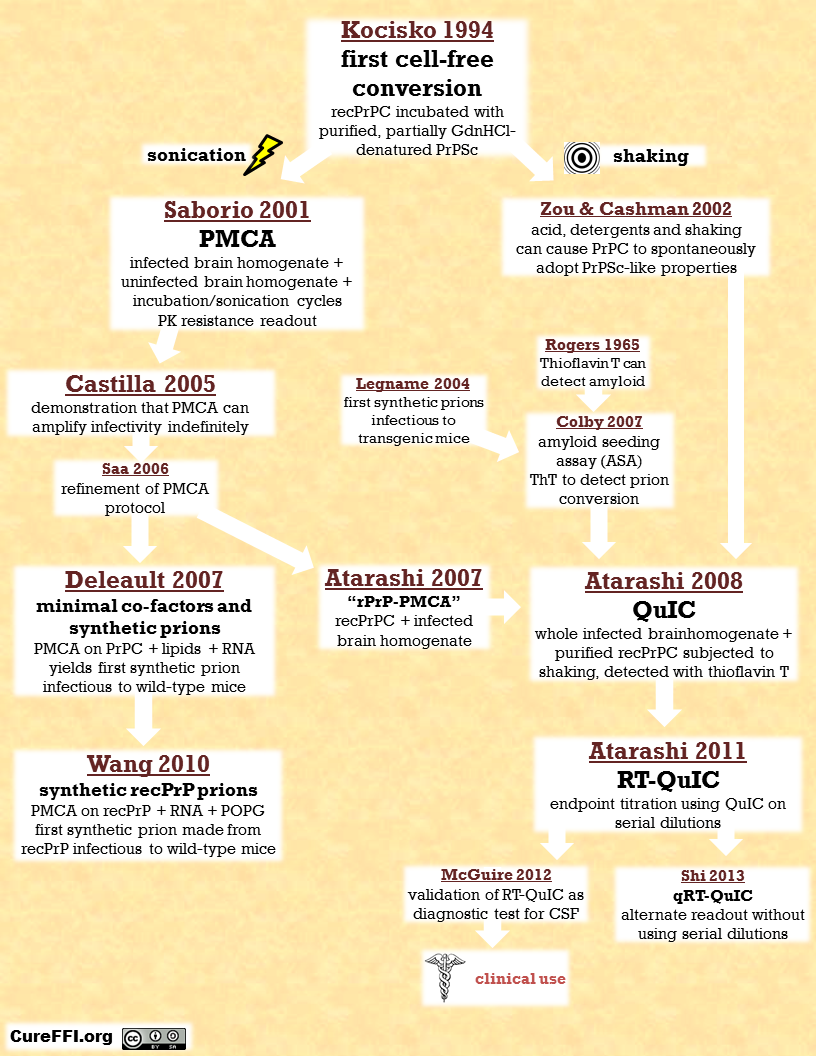
The evolution of cell-free prion conversion assays
Above is the best family tree I’ve been able to put together describing the evolution of assays stemming from the original discovery that PrPSc could convert PrPC in a cell free environment. In reality there have been far more papers than what I’ve included above, and I have probably neglected to include some things of great importance. Please leave a comment on this post to let me know how you’d structure the tree differently.
Here’s a more narrative look at how it all played out.
The story begins in the early 1990s with a bright, ambitious chemistry PhD student at MIT named Dave Kocisko. He worked in Peter Lansbury‘s lab and also worked closely with Byron Caughey at the NIH Rocky Mountain Labs in Montana. By then it was known PrPSc can be denatured (unfolded) by treatment with guanidine hydrochloride (GdnHCl). This group of researchers had noticed that if you treated PrPSc with highly concentrated (6M) GdnHCl, it lost its infectivity and its resistance to digestion by proteinase K (PK) for good. Even if you washed away the GdnHCl, it was too late: the PrP had already “forgotten” its scrapie conformation. But if you treated it with slightly less concentrated (3M) GdnHCl, it lost its scrapie properties only transiently: if you washed away the GdnHCl and let the PrP sit for a while, it would re-form into PrPSc fibrils, complete with PK resistance.
Their explanation for this phenomenon was as follows: 6M GdnHCl must completely denature all of the PrPSc, while 3M GdnHCl must just denature almost all of it, leaving behind some tiny crystal of PrPSc left that can serve to nucleate the polymerization of the rest of the PrP into scrapie fibrils. If that crystal could nucleate the denatured PrPSc to renature, they figured surely it could also nucleate fresh PrPC to join the nascent polymer as well. So Kocisko purified PrPSc, partially denatured it with 3M GdnHCl, and then mixed it with radiolabeled purified PrPC, and sure enough: some of the radiolabeled PrPC acquired PK resistance [Kocisko 1994].
For years, prions had been strictly an in vivo phenomenon [Prusiner 1982]. Prions had finally been successfully propagated in N2a cell cultures a few years earlier [Butler 1988 (ft)], but Kocisko’s experiments didn’t even involve cells – only purified protein. As such, this was the first real evidence for a specific mechanism of prion propagation: nucleated polymerization. The idea of this mechanism had been proposed even before Prusiner came onto the scene [Griffith 1967], but here at last was real evidence for it. In explaining it to the public, people occasionally make reference to zombies, but Byron Caughey, evidently a Kurt Vonnegut fan, saw the much better analogy to ice-nine [Lansbury & Caughey 1995].

Above: In Cat’s Cradle by Kurt Vonnegut, the U.S. military designs a new weapon, ice-nine, an alternate crystalline structure of water which is solid even at body temperature. One crystal of ice-nine, by providing a crystal seed that induces other water molecules to crystallize the same way, can cause a whole body of water to solidify.
Kocisko and others went on to do great things with this assay – for example, showing that the species barrier (now better described as a transmission barrier) in prion disease existed at the molecular level and must have to do with the ability of various PrP conformations to convert PrP molecules of unlike amino acid sequence [Kocisko 1995]. But it had a major limitation, which was that the PrPC conversion was substoichiometric. They put in 50 units of PrPSc and 1 unit of radiolabeled PrPC and got out < 1 unit of radiolabeled PrPSc. So this assay still couldn’t explain the apparently exponential growth of PrPSc in vivo.
Claudio Soto eventually had an insight that would fix this problem. Adding new PrPC monomers to polymers of PrPSc just gave you longer polymers, with probably linear growth, say, at both ends of a fibril. If you wanted exponential growth you had to shear up the big polymers into small oligomers that could each go out into the world and seed a new polymer of PrPSc. Once those got big enough, you had to shear them up again, and then repeat the whole process tens of times. Soto accomplished this with the intense ultrasonic noise of a sonicator. Thus was born protein misfolding cyclic amplification (PMCA) [Saborio 2001].
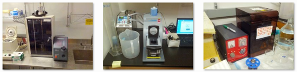
Above: a few of the various sonicators in our lab.
The original paper only showed that PMCA produced new PK-resistant PrP, but the authors went on to show that it also produced new infectivity. In fact, you could repeatedly amplify and dilute a sample so many times that there was less than a 1 in a million probability that even a single molecule of the original PrPSc seed was still present, and yet infectivity would still be present [Castilla 2005], although the PrPSc generated in vitro was quantitatively less infectious than bona fide in vivo prions (about 10-100 times less infectious per unit of protein). Using similar techniques, another group was able to generate PrPSc that did appear to be just as infectious as “real” prions [Bieschke 2004, Weber 2006].
Unlike Kocisko’s assay, which used purified protein, PMCA used whole brain homogenate from infected and uninfected animals. The PMCA reaction was cell-free in the sense that all the cells had been torn apart in a blender, but it did contain a lot else besides protein. This made it marginally harder to prove that the infectious agent you were generating was made of protein. Of course, there had already been an overwhelming body of evidence supporting the idea of infectious proteins for over 20 years at this point [Prusiner 1982], but there seems to have been a school of thought that the “final final final” proof of the prion hypothesis would be to generate an infectious prion from purified protein.
Toward that end, a recombinant mutant PrP which was simply incubated (but not sonicated) until it turned into a beta sheet rich form had recently proven infectious to transgenic mice overexpressing PrP [Legname 2004]. That was pretty convincing, but because the prions weren’t infectious directly to wild-type mice, it counted as perhaps only the “final final” proof and not the “final final final” proof. So people set out to see if PMCA could be adapted to demonstrate the generation of a prion from purified protein.
There were several efforts in this area, but I’ll mention a couple of the most notable. Nathan Deleault and Surachai Supattapone figured out that PMCA could work with just purified PrPC, PrPSc, RNA and lipids [Deleault 2007]. The lipids appeared to be 20-carbon fatty acids, and they weren’t added – they were just there because they couldn’t be separated from PrP. The sequence of the RNA didn’t seem to matter – in fact, any old single-stranded polyanion would do, and they just used polyA RNA. This new version of PMCA could amplify infectivity from a PrPSc seed, and in fact you could even do it without the seed and sometimes get a spontaneous prion to form which would be PK-resistant and infectious. Some people still would not accept this as the “final final final” proof, perhaps because the PrPC was brain-derived and not recombinant. So a few years later, Ma Jiyan and his PhD student Wang Fei screened a few other cofactors to see which had the best properties in PMCA, and settled on RNA and 1-palmitoyl-2-oleoylphosphatidylglycerol (POPG), a synthetic phospholipid. With just bacterially expressed, recombinant PrPC and these two molecules in PMCA, he created a de novo infectious prion [Wang 2010]. Only after they repeated this experiment in a brand new lab in Shanghai where no prion had ever been and there was no possibility of cross-contamination did people finally accept that this was indeed the “final final final” proof and give Wang Fei the Young Researcher Prize at Prion2013.
With that, hopefully the de novo prion issue can finally be put to bed. But meanwhile, loads of effort had also gone into a different problem, one of how to detect small amounts of PrPSc - say, for diagnostic purposes or to certify the safety of blood supplies. This spawned several efforts to adapt PMCA for highly sensitive prion detection, with some success [Saa 2006a, Castilla 2005b]. One hiccup was that PMCA used brain homogenate as a substrate – fine when you’re studying mice, but where were you going to get loads of human brain homogenate to do diagnostic tests on human prions? Perhaps brains from HuPrP mice would have sufficed; anyway, this problem was solved when PMCA was made to work on recombinant PrP as a substrate [Atarashi 2007].
But there were still several other problems. PMCA took a pretty long time – for instance, one protocol used 144 30-minute cycles (that’s 3 days) [Saa 2006a]. And once it was done, you had to do a Western blot to look for PK-resistant PrP. That’s no one’s favorite thing to do on an industrial scale. Last but not least, it was just plain hard to get PMCA to work. PMCA is a well-oiled machine in Claudio Soto’s lab in Houston, but many other researchers describe PMCA as being finnicky at best. As Ryuichiro Atarashi put it:
With sonication of PMCA reaction tubes in cuphorn probes, the delivery of vibrational energy to samples can vary substantially with tube position, tube construction, probe age, bath volume, and the redistribution of samples within the tubes by sonication-induced atomization and condensation. [Atarashi 2008 - supplement].
So Atarashi and Byron Caughey invented a new assay, drawing on a couple different sources of inspiration. Instead of brain homogenate they used recombinant PrPC as the substrate, as they’d done in their adapted version of PMCA [Atarashi 2007]. They also substituted shaking for sonication, an idea they got from Zou & Cashman 2002. Whereas the energy of sonication was distributed unevenly (see above quote), shaking was a pretty uniform and predictable process. It’s amazing to me that this actually works. Sonication is high-energy stuff that can shear DNA and disrupt cells – pregnant women are forbidden to operate the sonicator in my lab. “Shaking” has a quality of vibration more like driving on a poorly maintained road or sitting in a bad massage chair at the mall.
Above: the shaker machine in our lab
But it worked. The new assay didn’t seem to produce any infectivity, but after enough rounds of shaking and incubation you reliably got a PK-resistant PrP amyloid which you could identify in a Western blot [Atarashi 2008].
Still, few things could be slower and more tedious than making a Western blot, so it took one final innovation to make the new assay into something truly powerful. They soon found that instead of blotting for PK resistance they could use Thioflavin T (ThT) – a compound that fluoresces in the presence of beta sheet rich proteins and thus can detect amyloid. That idea had been around a good long while [Rogers 1965], and the Prusiner lab had adapted it for a new so-called amyloid seeding assay (ASA) [Colby 2007]. With ThT, you just needed to put the 96-well plate in a platereader and watch for fluorescence.
To adapt this new assay to precisely quantifying the amount of PrPSc present in a sample, they then borrowed a very old trick. Back in the “scrapie agent” days people quantified the “agent” by doing serial dilutions – 10-1, 10-2, 10-3 etc., and injecting each dilution into a separate group of mice. So if for instance the first dilution where not all animals died was 10-9, then you figured your sample had ~109 LD50 units in it. This was called the Spearman-Karber method; people cite Dougherty 1964. Prusiner more or less put an end to this time-, space- and mouse-intensive nonsense when he showed that you could just inoculate one group of animals with one diultion and that the incubation time was correlated with the prion titer (this is called the incubation time bioassay) [Prusiner 1982b]. Still, people do occasionally bust out the old Spearman-Karber as a gold standard when they want to show that their new assay is just as good [Tamguney 2009].
By analogy to the Spearman-Karber bioassay, Atarashi and Caughey performed RT-QuIC on serial dilutions, and if, say, 10-9 was the first dilution that didn’t reach ThT fluorescence saturation after 48 hours of cycling, then you had 109 SD50s in your sample. Not LD50 – because we’re not measuring lethality – but SD50, the median seeding dose. This new assay – QuIC with serial dilutions – was dubbed RT-QuIC, by analogy to RT-PCR (better known as qPCR) [Atarashi 2011]. People are still tinkering with this assay – for instance, this year a new study uses the time to ThT saturation at one dilution, rather than the minimum dilution to reach saturation, as the assay readout and calls this qRT-QuIC [Shi 2013]. qRT-QuIC is to QuIC as incubation time bioassay is to Spearman-Karber method.
A blind screen of > 200 cerebrospinal fluid samples from humans showed that RT-QuIC had ~90% sensitivity and ~100% specificity as a prion disease diagnostic tool, at least for sporadic Creutzfeldt-Jakob disease [McGuire 2012]. Another (though smaller) screen suggested similar levels of accuracy for several forms of genetic prion disease, including fatal familial insomnia [Sano 2013]. That’s good news because other sCJD tests such as diffusion-weighted MRI [Vitali 2011] don’t seem to work quite as well on other prion strains (see CJD2013 highlights).
So RT-QuIC seems to be the new biochemical prion diagnostic tool we’ve been needing. At Prion2013 Valerie Sim told me that it was about to reach the clinic in Canada and that she would probably be sending patient CSF samples off to be tested within a couple of months. I haven’t heard an update on this and when I Googled I didn’t find any news articles about it yet. But the consensus at Prion2013 seemed to be that clinical use was just around the corner. Hopefully better, earlier diagnosis means earlier intervention in the next clinical trial of a treatment.
Looking forward, another hopeful area will be to determine whether any of these assays -QuIC, PMCA or something in between – can predict the strain specificity of different therapeutic molecules, a question I discussed at the end of this post. For instance, it was shown that anle138b inhibited the PMCA reaction for a wide range of different prion strains [Wagner 2013], and if this indeed turns out to be predictive of the drug’s efficacy against those strains in vivo, then years could be shaved off of drug development timelines by not bothering to do lead development and pharmacokinetics on drugs that don’t work on human prion strains.

About Eric Vallabh Minikel
Eric Vallabh Minikel is on a lifelong quest to prevent prion disease. He is a scientist based at the Broad Institute of MIT and Harvard.