Robert Thorne: blood-brain barrier primer
Dr. Robert Thorne (Denali Therapeutics; University of Minnesota; co-founder of International Brain Barriers Society) visited our lab today to give us a primer on the blood-brain barrier: “Engineering delivery of antibodies, enzymes, & oligonucleotides to the brain & other body tissues through transferrin receptor targeting: transporting biotherapeutics to the final frontier.” These are my notes.
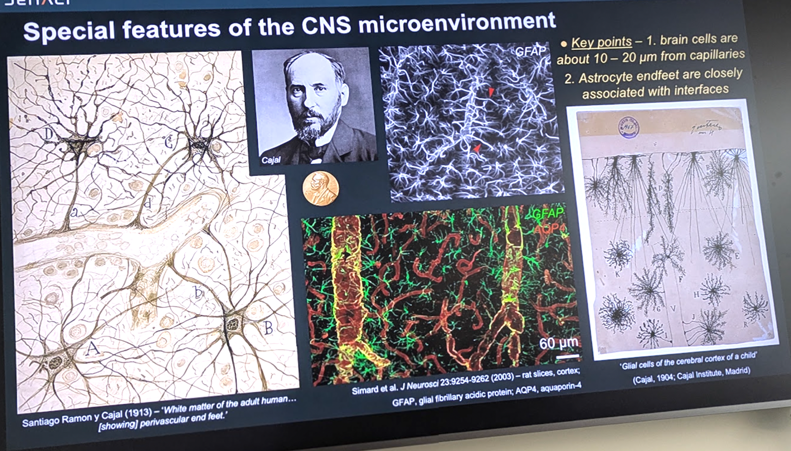
Santiago Ramon y Cajal, winner of the first neuroscience-related Nobel Prize, did a lot of staining to illuminate structures of the CNS. He was among the first to observe the endfeet of astrocytes arrayed at the border of capillaries and other blood vessels in the brain (shown in the image is a likely arteriole or venule based on vessel diameter). All neurons and glia are within about 10 - 20 μm of capillaries, and this distance is preserved across species because it is likely constrained by the limits on thef diffusion of things such as oxygen, glucose and other essential nutrients. Astrocyte endfeet are closely associated with fluid interfaces.
There are 5 FDA-approved CNS therapies delivered either intrathecally or intraparenchymally: ziconotide (intrathecal; chronic pain), nusinersen (intrathecal; SMA), cerliponase alfa (intraventricular; Batten), tofersen (intrathecal; SOD1 ALS), and AAV2:AADC (intraparenchymal injection into striatum; aromatic amino acid decarboxylase deficiency). There are also 5 FDA-approved therapies non-invasively delivered to the brain intravenously: aducanumab, lecanemab, and donanemab (mAbs for Alzheimer’s), onasemnogene abeparvovec (AAV SMN1 for SMA), and tividenofusp alfa (Denali’s new TfR-targeted protein therapeutic for Hunter syndrome).
There have been 2 decades of progress in AAV delivery to the CNS. Kaspar and colleagues observed that IV AAV9 worked better with respect to transducing neurons in neonatal mice, whereas it transduction was much more limited to astrocytes in adult mice [Faust 2009]. The volume of intracellular space at birth is 40% of total brain volume, whereas in adulthood it drops to 20% because oligodendrocytes, astrocytes and other cells proliferate and mature and fill in the remaining space. Intracisterna magna (ICM) injection of AAV is constrained by AAV size, ~25 nm diameter. Extracellular space has a very conserved dimension that limits the diffusion distance, so brain coverage does not scale with brain size. That diffusion is already fairly limited and uneven in mouse brain [Bey 2017] and is even more limited to cells at the surface of the cortex in monkeys due to larger brain size [Samaranch 2014]. Oligonucleotides are far smaller than AAV, and you can calculate diffusion coefficients, but you still would not expect a ton of deep brain access from intrathecal administration. The extracellular space was long thought to be 10 - 20 nm by EM imaging. And yet extracellular space is about 20% of total brain volume across humans and a wide range of animal species, which cannot be explained by such narrow extracellular space. Using a range of molecular weights of dextran and PEGylated quantum dots, they looked at diffusion of particles ranging 3 nm to 35 nm in adult rats [Thorne & Nicholson 2006]. This determined the size of the extracellular space was approximately 40 to 60 nm, depending whether you model it as parallel planes or cylindrical pores. Let’s say it’s roughly 50 nm. This gives you some kind of constraints on what kind of macromolecules might be able to diffuse at all.
The brain lacks conventional lymphatics; there is some hypothesis that these perivascular spaces — on the CNS side of the BBB, along blood vessels — function as a glymphatic system, but the topic remains controversial in the field [Abbott 2018].
In peripheral organs, endothelial cells that line the blood vessels have larger fenestrations that allow macromolecules to flow from blood into the organ. These are largest in liver — 150 nm diameter, which is why everything goes to the liver. It is 20-30 nm in kidney, and 6-7 nm in skeletal muscle, skin, and mucus membranes. The vessels in these tissues have very little expression of transporters or receptors such as GLUT1 or TfR. The brain is opposite: endothelial cells form tight junctions that allow no passive paracellular diffusion into the tissue, and instead, there is high expression of GLUT1 and TfR. Pericytes are thought to have a major role in development of the BBB during embryogenesis in humans.
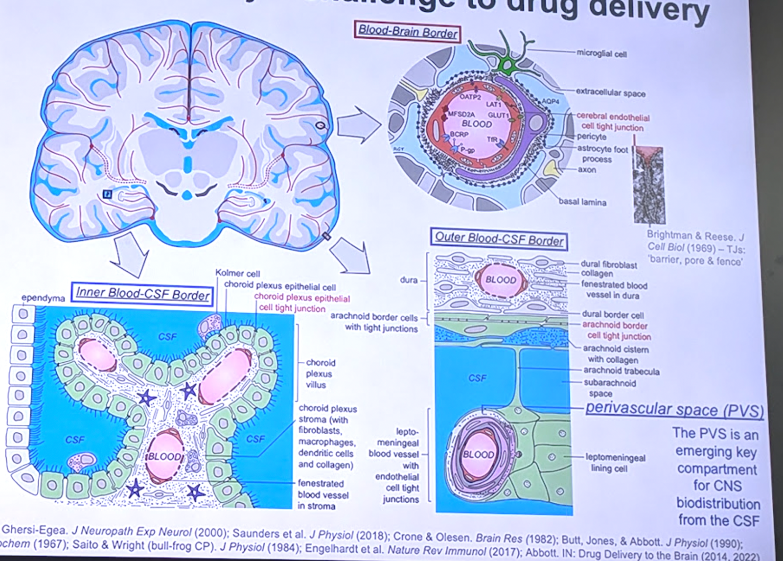
Thorne and colleagues have likened the BBB to a border between countries [Badaut 2024] — it is a complex, dynamically regulated interface that has to let some things through, but is not simply wide open to everything. The neurovascular unit view of things is that every neuron or glial cell has a source of blood within 10-20 μm. There are different morphologies of the blood-brain barrier inner blood-CSF border (choroid plexus; deep inside the brain within the ventricles) versus the outer blood-CSF border (at the surface of the brain, along the arachnoid border).
Joan Abbott very elegantly diagrammed out the different ways that things get into the brain [Abbott 2006]:
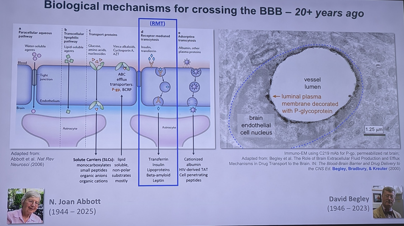
Begley showed that in brain endothelial cells, some of the relevant proteins can be highly polarized meaning they are almost exclusively on either the blood side or the brain side, but not both. A challenge in studying these endothelial cells is that they get very thin as they wrap around the vessel lumen, below the resolution of light microscopes. Chenghua Gu’s lab has focused on the side of the endothelial cell that contains the nucleus, which forces it to be thicker, and enables quantifying which side of the cell the different proteins are on — for instance, P-gp is almost exclusively luminal, while GLUT1 is about 50/50 [Amick 2026].
8 targets that have some preclinical or clinical data showing they can be leveraged for trans-BBB transport are TfR, INSR, IGF1R, LRP1, ADAM15, ALPL, CA4, and CD98hc. Some of these are “zonated” meaning they are preferentially expressed in more venous or more arterial blood vessels, for example, TfR is expressed in microvessels (capillaries and postcap venules) while having low expression in arteries and arterioles [Vanlandewijck 2018, Yang 2022]. A lot of these proteins including TfR have stable expression across age groups in humans, but substantial inter-individual variation [Santa-Maria 2026]. But protein abundance alone may not explain transport efficiency — there could also be factors affecting luminal sorting, flux rate, etc. Indeed, some recent data show that in mice, at least, the amount of TfR-shuttled IgG that enters the brain is higher in neonates and then declines to a stable adult level that is maintained even in very aged mice [Torres 2026]. Mannose-6 phosphate receptor (M6PR; IGF2R) is even worse, it’s expressed on the BBB only through 2-3 weeks of age in mice and then is pretty much absent.
That was all just the introduction. We are now at the cusp of being able to leverage all this to engineer drugs to get into the brain [Abbott 2025, Dolgin 2026]. Denali engineered transport vehicles (TV) with a 9 amino acid patch to cross the BBB by targeting human TfR [Kariolis 2020] or human Cd98hc [Chew 2023]. This was done by screening a bunch of different amino acid sequences and then using affinity maturation and other methods to optimize binding and cross species affinity. They got ones with a wide range of affinity values. TfR has higher capacity and peaks very quickly, in about 1 day; Cd98 peaks in 5-7 days. They can also be used in combination [Wells 2025].
TfR’s highest expression is on erythroid progenitors in the bone marrow and also on reticulocytes (immature red blood cells) so one of the biggest challenges for TfR-targeted therapeutics is anemia. The 2nd highest is BBB endothelial cells, and 3rd highest is neurons. Cd98 has a different expression profile which helps diversify it. The receptors have distinct distribution profiles across both brain and peripheral tissues [Khoury 2025]. Interestingly, IgG that lacks a BBB-targeting domain, to the extent that it enters the brain at all, appears to do so by entering CSF rather than crossing BBB, and so is actually limited by the same CSF diffusion patterns you would see with CSF delivery [Khoury 2025, Hohsfield 2025].
Each TfR molecule binds to 2 ferric iron (Fe3+) ions. On the other hand, ferritin, the major intracellular iron binding protein that is also present in low levels extracellularly, binds 4,500 Fe3+ in a big cage. Its transcytosis is tightly regulated to make sure those iron ions are not suddenly released into the endothelial cell. Denali’s TV platform monovalently targets TfR. The TV can work as an enzyme or protein fusion, or with a linker to an oligonucleotide.
For using the TV for oligo delivery, they compared intrathecal vs. BBB-shuttled MALAT1 ASO in cynomolgus macaques [Barker 2024]. The intrathecal ASO was most intense at the surface of cortex, with little diffusion into deeper brain, while the BBB-shuttled one was more uniform throughout brain. They now have a shuttled MAPT oligonucleotide in a healthy volunteer trial. They are also using the TV to deliver an anti-amyloid antibody [Pizzo 2025]. It is an asymmetric antibody where they introduced the TfR-binding TV and a “LALA patch” on just 1 side of the antibody, to prevent simultaneous binding of Fc gamma receptors and TfR. They do want some degree of effector function, but only when the Fabs are bound to amyloid in brain and not when the TV is bound to TfR onin reticulocytes. The distribution of a shuttled anti-amyloid antibody is expected to not result in as much exposure in perivascular space as a non-shuttled antibody, and thus, may reduce ARIA, the main side effect of anti-amyloid antibodies [Pizzo 2025, Alzforum post]. Human autopsy data suggest that aducanumab target engagement is mostly in very superficial layers of cortex, even in patients where PET scans suggested more profound amyloid lowering [Boon 2025]. Florbetapir, the PET tracer, is a small enough molecule that it probably diffuses uniformly through brain after crossing the BBB, but it’s not clear if it binds all forms of amyloid.
Wilhelm His in 1865 already hypothesized that the perivascular spaces of the CNS had a lymphatic-like function. In a study of intrathecal antibody distribution pathways in rat, Pizzo et al modeled out the diffusion coefficients for IgG (150 kDa) vs. single-domain antibody (sdAb; 15 kDa) [Pizzo 2018]. This is not an artifact of ex vivo tissue processing, it has replicated with gadolinium labeling and in vivo imaging.
Denali’s Hunter syndrome (MPS II) drug, tividenofusp alfa, uses Denali’s TV to transport the enzyme into the brain while also reaching peripheral tissues [Ullman 2025, Bhalla 2020, Arguello & Mahon 2022]. It received FDA Accelerated Approval in March 2026. The clinical dose is 15 mg/kg once weekly. It leverages TfR to cross the BBB and M6PR to enter neurons and glia where it stays in lysosomes and degrades glycosaminoglycans. In the clinical trial of 47 boys, infusion-related reactions, a known risk of ERTs, were the most common adverse event, decreasing in incidence and severity over time while treatment was associated with reductions in CNS and peripheral biomarkers of substrate accumulation and neuronal injury to levels within the range of unaffected children [Muenzer 2026]. Heparan sulfate in CSF and in urine got below the upper limit of normal pretty quickly. Serum NfL levels appeared to be reduced from baseline by 21% at week 49 and by 76% at week 153, with 85% of the participants having levels in the range of healthy children at week 153 [Muenzer 2026].

About Eric Vallabh Minikel
Eric Vallabh Minikel is on a lifelong quest to prevent prion disease. He is a scientist based at the Broad Institute of MIT and Harvard.